
Title: "A Comprehensive Review of Developments in Neuroimaging Technology"
Exploring the Frontiers of Neuroimaging: A Comprehensive Review of Cutting-Edge Technologies and Innovations

Neuroimaging is a term for medical imaging techniques used to investigate brain anatomy, function, and pathology. In this comprehensive overview, we will look at how neuroimaging can be used to diagnose and manage a variety of brain-related conditions, including neurodegenerative disorders, brain tumours, stroke, traumatic brain injury, and more. We examine the most recent breakthroughs in neuroimaging modalities, such as magnetic resonance imaging (MRI), positron emission tomography (PET), and functional magnetic resonance imaging (fMRI), emphasizing their roles in illness diagnosis, treatment planning, and prognosis. Furthermore, we investigate the transformative impact of artificial intelligence (AI) and machine learning (ML) approaches on neuroimaging data, namely in disease diagnosis and prognosis prediction. Notably, we address new findings that show the effectiveness of deep learning algorithms in detecting small irregularities associated with neurological disorders such as Alzheimer's disease and Parkinson's disease. Furthermore, we investigate the impact of brain-computer interfaces (BCIs) in restoring motor function and improving cognitive capacities by combining ML and DL approaches. Despite major advances, difficulties remain, such as imaging protocol standardization and biomarker validation. Future research directions include multimodal imaging methods, large-scale data analytics, and precision medicine programs aimed at improving patient outcomes and expanding our understanding of brain-related disorders. Collaboration between researchers, physicians, and industry stakeholders is critical for driving innovation and implementing cutting-edge imaging technologies in clinical practice, ultimately improving patient care and quality of life.
Neuroimaging, often known as BRAIN Imaging, is a technology that uses multiple imaging techniques to capture the anatomy of the brain or other parts of the nervous system. Each neuroimaging approach has its own method for taking visual images of the brain and assisting healthcare providers in diagnosing neurological problems. Different approaches are used to diagnose and confirm certain neurological illnesses. Brain-related diseases pose significant challenges to healthcare systems worldwide, necessitating accurate and timely diagnosis for effective management. The doctors employ a variety of parameters in addition to the results from the neuroimaging modalities to determine the disease; nonetheless, the neuroimaging data plays an important role in validating and assisting the doctors in reaching judgments.
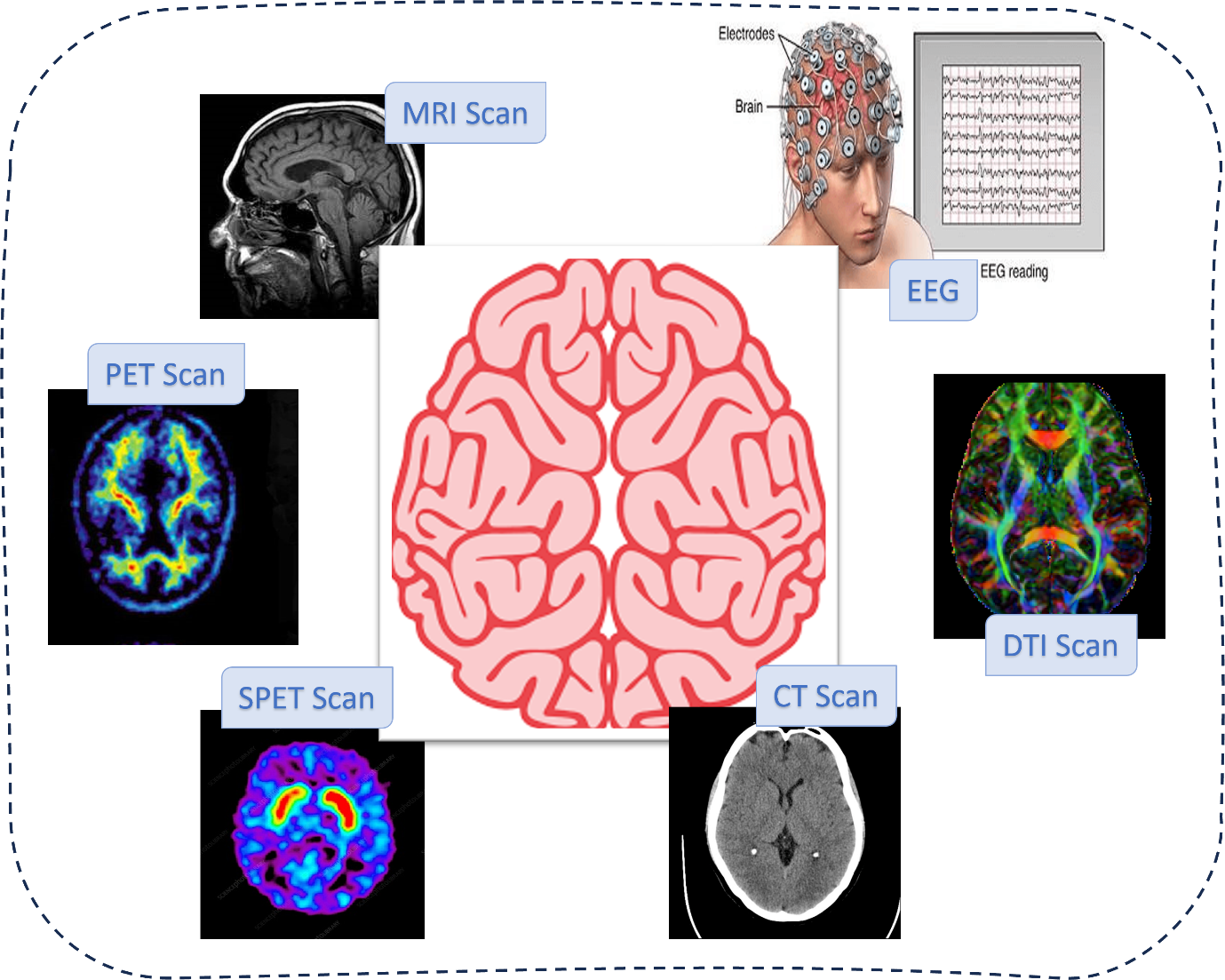
There are different medical imaging techniques like magnetic resonance imaging (MRI), computed tomography (CT), positron emission tomography (PET), and single-photon emission computed tomography (SPECT) that have transformed our understanding of brain anatomy, function, and pathology. The purpose of this study is to look at a Comprehensive Review of Developments in Neuroimaging Technology and how they might be used to diagnose and manage brain illnesses. In table 1, a few neuroimaging techniques are listed. A graphical representation of each neuroimaging modality can be visualized in figure 1. MRI scan is a 3D scan for 3 views of brain; axial, coronal and sagittal. It creates detailed images of the brain and other body areas by combining a powerful magnetic field and radio waves. Similarly, CT scans employ X-rays to provide detailed images of the brain and other organs. It is commonly used to diagnose brain ailments such as migraines and haemorrhages. EEG measures the functioning of the brain by monitoring its electrical activity using electrodes put on the top of the head. It is frequently used to identify epilepsy and other conditions that affect cognitive function.
| Neuroimaging Techniques |
| Magnetic Resonance Imaging (MRI) |
| Functional magnetic resonance imaging [fMRI] |
| Positron Emission Tomography (PET) |
| Magnetoencephalography [MEG] |
| Single-photon emission computed tomography [SPECT] |
| Electroencephalography [EEG] |
| In vivo magnetic resonance spectroscopy |
| Near-infrared spectroscopy [NIRS] |
| Functional near-infrared spectroscopy |
| Magnetic resonance imaging of the brain |
| Diffusion Tensor Imaging [DTI] |
| Diffuse Optical Imaging |
| Cranial ultrasound |
| Computerised Tomography (CT) |
| Optical coherence tomography (OCT) |

Figure 1: Different Neuroimaging Techniques used to capture brain functions and structure.
Table 4 summarizes the data type, nature of the data, and the principal imaging technology used to capture the data or the method on which the data is based from the brain. The findings aid in the planning of preprocessing procedures and the processing of data for the use and study of ML and DL technologies.
| Neuroimaging technique | Data generated | Technique data is based on |
| MRI | Images | Captures pathologies, tissue properties, brain activity and blood flow velocity. |
| fMRI | Images | Brain activity by detecting changes in blood flow. |
| PET | Images | The metabolic or biomedical function of tissues. |
| EEG | Signals | Recording of the electrical activity of the brain from the scalp. |
| CT | Images | X-Ray images are taken from different angles and create cross-sectional images. |
| SPECT | Images | A nuclear imaging test using a radioactive substance and a special camera to create a 3D picture. |
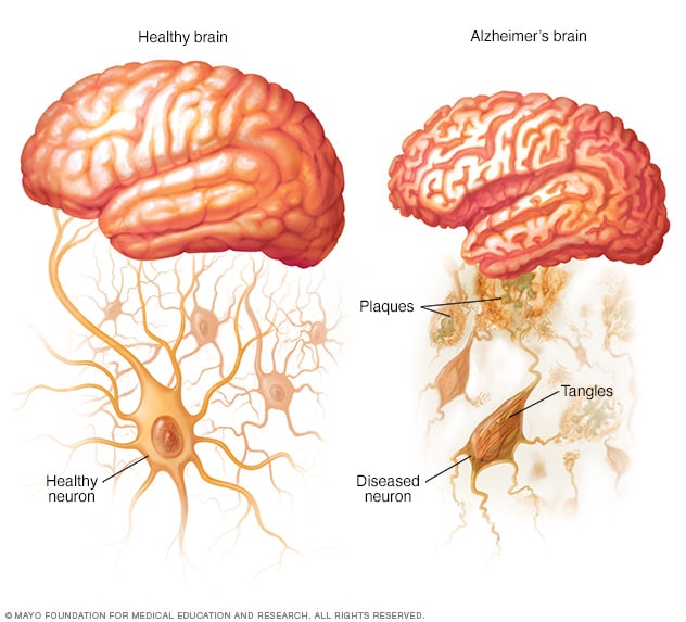
Alzheimer's disease, Parkinson's disease, and multiple sclerosis are all neurodegenerative disorders that cause progressive neuronal degeneration and associated cognitive or motor issues. Advanced MRI techniques, such as diffusion tensor imaging (DTI), functional MRI (fMRI), and magnetic resonance spectroscopy (MRS), provide important insights into the anatomical and functional alterations in the brain associated with these illnesses. Furthermore, molecular imaging technologies such as PET and SPECT aid in the early detection of pathogenic biomarkers, allowing doctors to undertake prompt therapies and track disease development. Recently, many studies are conducted for efficient classification and disease progression detection for as Alzheimer's disease [1], Parkinson's disease [2], and multiple sclerosis [3].

Figure 2: Normal Brain and Alzheimer’s Brain.
Neuroimaging plays a crucial role in the diagnosis, characterization, and treatment planning of brain tumours. High-resolution MRI with contrast enhancement remains the cornerstone for evaluating tumour location, size, and morphology. Advanced neuroimaging modalities, such as perfusion-weighted imaging (PWI), magnetic resonance spectroscopy (MRS), and diffusion-weighted imaging (DWI), provide valuable insights into tumor blood flow, metabolic function, and tissue density, which help distinguish between different conditions and evaluate treatment effectiveness. Moreover, PET imaging with radiotracers targeting specific molecular pathways offers valuable insights into tumour biology and treatment planning, particularly in gliomas and metastatic brain tumours [6].
Neuroimaging plays a critical role in the acute management and long-term monitoring of stroke and cerebrovascular diseases. CT and MRI are routinely used to assess the extent of cerebral ischemia, identify underlying etiology, and guide therapeutic interventions such as thrombolysis and endovascular clot retrieval. Advanced imaging techniques such as perfusion imaging, diffusion-weighted imaging, and magnetic resonance angiography (MRA) enable clinicians to evaluate tissue viability, assess collateral circulation, and predict clinical outcomes following stroke. Furthermore, emerging imaging modalities such as functional MRI and resting-state fMRI hold promise for mapping neuroplasticity and predicting recovery trajectories in stroke patients.
Neuroimaging plays a crucial role in the diagnosis and management of traumatic brain injury, facilitating the assessment of structural damage, intracranial hemorrhage, and associated complications. CT remains the primary imaging modality for the initial evaluation of TBI due to its widespread availability and rapid image acquisition. Advanced MRI techniques, including susceptibility-weighted imaging (SWI) and diffusion tensor imaging (DTI), provide valuable insights into the microstructural and functional alterations in the brain following TBI, aiding in prognostication and treatment planning. Moreover, emerging imaging biomarkers such as tau PET imaging hold promise for detecting chronic traumatic encephalopathy (CTE) and monitoring disease progression in athletes and individuals exposed to repetitive head trauma.
In the realm of neuroscience, the exploration of the human brain's intricacies relies heavily on the insights provided by neuroimaging. While neuroimaging itself isn't an aspect of artificial intelligence (AI), the marriage of AI techniques, particularly machine learning (ML) and deep learning (DL), has significantly enriched this field.
Neuroimaging techniques like functional magnetic resonance imaging (fMRI), positron emission tomography (PET), and diffusion tensor imaging (DTI) generate extensive datasets containing intricate information about brain structure, function, and connectivity. However, extracting meaningful insights from such complex data requires sophisticated analytical methods due to the multidimensional and high-dimensional nature of neuroimaging data.
Machine learning (ML) algorithms emerge as powerful tools in this context, capable of effectively recognizing patterns and extracting valuable information from large and intricate datasets. ML techniques, including supervised learning, unsupervised learning, and reinforcement learning, are adept at handling the complexities and nuances present in neuroimaging data.
For instance, in functional MRI (fMRI) data analysis, ML algorithms can identify brain regions that exhibit synchronized activity patterns, revealing functional connectivity networks associated with specific cognitive tasks or neurological conditions. Similarly, in PET imaging, ML algorithms can detect and quantify abnormal metabolic patterns indicative of neurodegenerative diseases or tumors, aiding in early diagnosis and treatment planning.
Moreover, diffusion tensor imaging (DTI) captures the diffusion of water molecules in brain tissue, providing insights into white matter microstructure and connectivity. ML algorithms can analyze DTI data to reconstruct white matter pathways, detect abnormalities in fiber tracts, and investigate alterations in brain connectivity associated with neurological disorders.
A notable example is the work by Yeo et al. [7], published in the journal Nature Neuroscience, where ML techniques were employed to identify distinct functional connectivity patterns in the human brain based on resting-state fMRI data. The study demonstrated the ability of ML algorithms to uncover complex brain networks and their relevance to cognitive functions and neurological disorders.
In summary, ML algorithms play a crucial role in neuroimaging data analysis by enabling the extraction of meaningful insights and patterns from complex datasets. Their ability to uncover subtle neurological phenomena holds great promise for advancing our understanding of brain function and dysfunction, ultimately leading to improved diagnosis, treatment, and management of neurological disorders.
One of the most promising applications of machine learning (ML) and deep learning (DL) in neuroimaging lies in disease diagnosis and prognosis. By leveraging ML and DL techniques on large datasets of brain scans from both healthy individuals and patients with neurological disorders, such as Alzheimer's disease, Parkinson's disease, or schizophrenia, researchers can develop highly accurate models capable of detecting subtle abnormalities indicative of these conditions. These algorithms analyze complex patterns and features within neuroimaging data, providing valuable insights into the structural and functional alterations associated with various neurological disorders.
For instance, in a study published in the journal Nature Medicine titled "Deep Learning Predicts Brain Age and Alzheimer's Disease Using Resting-State fMRI," Cole et al. [5] demonstrated the efficacy of deep learning algorithms in predicting brain age and diagnosing Alzheimer's disease using resting-state functional magnetic resonance imaging (fMRI) data. The study showcased the potential of DL models to accurately identify neurological abnormalities associated with Alzheimer's disease, enabling early diagnosis and intervention.
Moreover, ML algorithms play a crucial role in predicting disease progression and treatment outcomes, thereby facilitating personalized healthcare approaches tailored to each patient's unique neurological profile. By analyzing longitudinal neuroimaging data and clinical information, ML models can forecast the trajectory of neurological diseases, anticipate changes in cognitive function, and optimize treatment strategies.
A notable example is the work by Khosravi et al. [6], published in the Journal of Neuroscience Methods, where ML algorithms were employed to predict the progression of Parkinson's disease based on multimodal neuroimaging data. The study demonstrated the potential of ML-based prognostic models in guiding clinical decision-making and improving patient care.
In summary, ML and DL techniques hold tremendous promise in neuroimaging for disease diagnosis and prognosis, enabling earlier detection, personalized treatment planning, and improved patient outcomes. These advancements pave the way for more effective management of neurological disorders and highlight the transformative potential of AI-driven approaches in neuroscience research and clinical practice.
ML and DL techniques are also driving advancements in brain-computer interfaces (BCIs), which establish direct communication pathways between the brain and external devices. By decoding neural activity captured through neuroimaging technologies, BCIs hold the potential to restore motor function to individuals with paralysis, enhance cognitive abilities, and even enable novel forms of human-computer interaction. ML algorithms play a crucial role in translating raw brain signals into actionable commands, paving the way for more intuitive and efficient BCIs.
Despite the significant advancements in medical imaging/neuroimaging for brain-related diseases, several challenges remain, including the need for standardized imaging protocols, validation of imaging biomarkers, and integration into clinical practice. Furthermore, the emergence of artificial intelligence (AI) and machine learning (ML) algorithms holds promise for automated image analysis, personalized risk prediction, and treatment optimization in brain disorders. Future research efforts should focus on harnessing the synergistic potential of multimodal imaging approaches, large-scale data analytics, and precision medicine initiatives to improve patient outcomes and advance our understanding of brain-related diseases.
Neuroimaging plays a central role in the diagnosis, treatment, and management of brain-related diseases, offering invaluable insights into brain anatomy, function, and pathology. By leveraging the latest advancements in imaging technology and computational methods, clinicians can achieve earlier and more accurate diagnoses, personalize treatment strategies, and monitor disease progression over time. As discussed previously different neuroimaging modalities, such as magnetic resonance imaging (MRI), positron emission tomography (PET), and functional magnetic resonance imaging (fMRI), have played important role in disease diagnosis, treatment planning, and prognosis. Moreover, there are state-of-the art methods of machine learning and deep learning that give promising results of disease detection and diagnosis using different neuroimaging modalities discussed in this article. These methods have revolutionized the traditional disease diagnostic procedures by providing non-invasive procedure to diagnose and monitor various neurological conditions. Continued collaboration between researchers, clinicians, and industry stakeholders is essential to drive innovation, overcome existing challenges, and translate cutting-edge imaging technologies into clinical practice, ultimately improving patient outcomes and quality of life for individuals affected by brain-related diseases.