

Brain tumors affect over 308,000 people globally each year, with an estimated 252,000 new cases diagnosed annually. Of these, nearly 60% are malignant, contributing to over 240,000 deaths worldwide. Early detection is vital, as studies show that timely intervention can increase survival rates by up to 50%, while late diagnoses often lead to more aggressive treatments and lower survival rates. Traditional diagnostic methods like MRI and CT scans can take hours to interpret and are susceptible to human error, with misdiagnosis rates reaching up to 15%. AI-powered systems, particularly Convolutional Neural Networks (CNNs), have shown the potential to reduce diagnostic time by up to 60% and improve accuracy by over 90%. By integrating AI into brain tumor classification, healthcare systems can significantly enhance early detection, leading to improved patient outcomes and more personalized, effective treatments.
The classification of brain tumors is a critical task in medical imaging and healthcare, where early detection can significantly improve patient outcomes. With the rise of artificial intelligence (AI) and machine learning (ML) technologies, the process of diagnosing brain tumors has become more accurate and efficient. This blog delves into the advancements in brain tumor classification, the challenges faced, and the role of AI in revolutionizing this field.
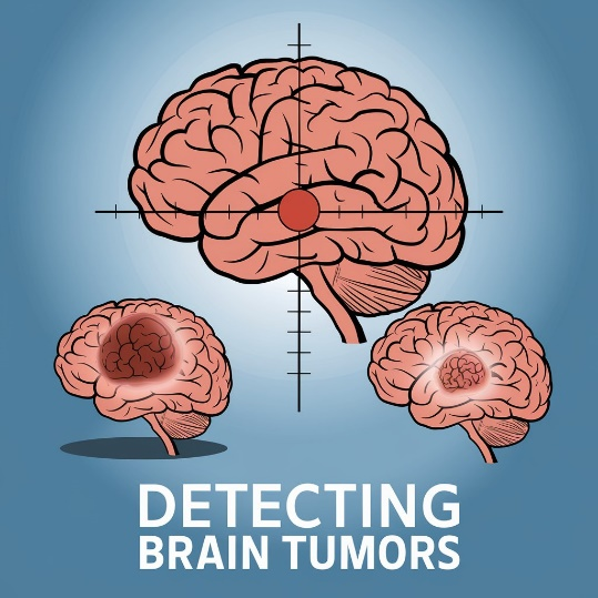
Brain tumors are abnormal growths of cells in or around the brain. These tumors can either be benign (non-cancerous) or malignant (cancerous). Based on their origin, brain tumors are classified into two categories:
Primary Tumors: Originate in the brain.
Secondary (Metastatic) Tumors: Originate elsewhere in the body and spread to the brain.
Early detection of brain tumors is crucial because it can lead to more effective treatment options, improve survival rates, and potentially reduce the invasiveness of surgical procedures. Brain tumors can grow rapidly, and without timely intervention, they can cause significant damage to the brain, leading to neurological impairments or even becoming life-threatening.
Traditionally, diagnosing brain tumors relies on medical imaging techniques such as Magnetic Resonance Imaging (MRI), Computed Tomography (CT) scans, and biopsy procedures. MRI is considered the gold standard due to its ability to provide high-resolution images of soft tissues, allowing clinicians to detect abnormalities. However, this manual process presents several challenges:
Time-Consuming: Physicians and radiologists must carefully examine numerous MRI slices and consider various clinical factors, making the process lengthy.
Complexity of Brain Structures: The human brain has complex structures and identifying subtle differences between healthy and tumor-affected tissue can be difficult, especially in the early stages.
Prone to Human Error: Manual inspection of images can be influenced by fatigue, variability in expertise, and even cognitive biases. Misdiagnosis or delayed diagnosis can result in poor treatment outcomes.
Artificial intelligence can address many of these limitations by providing a more efficient and accurate diagnostic process:
Speed and Efficiency: AI models can analyze MRI scans much faster than humans, processing thousands of images in a fraction of the time. This can significantly reduce the time required for diagnosis, enabling quicker treatment decisions.
Improved Accuracy: AI models, particularly deep learning techniques like Convolutional Neural Networks (CNNs), are trained on large datasets of MRI images and can detect minute patterns or anomalies that might be overlooked by a human expert. These models can differentiate between tumor types, identify tumors in their early stages, and even suggest the likelihood of malignancy with high precision.
Reduced Variability: Since AI models are consistent and unaffected by external factors like fatigue or cognitive bias, they reduce the risk of variability in diagnosis. This means that AI-assisted diagnostic tools can produce more reliable results, ensuring that early signs of brain tumors are not missed.
Supporting Radiologists: Rather than replacing doctors, AI can act as a decision support tool, assisting radiologists in making faster, more accurate diagnoses. By flagging potential tumor areas or generating preliminary reports, AI allows medical professionals to focus on more complex cases and make informed treatment decisions.

When brain tumors are detected early:
Less Aggressive Treatment: In many cases, early-stage tumors can be treated with less aggressive approaches, such as targeted therapies or minimally invasive surgeries, which reduce risks and complications.
Better Prognosis: Early detection often correlates with a higher survival rate and better quality of life post-treatment, as doctors can intervene before the tumor progresses to an advanced stage.
Lower Healthcare Costs: Early diagnosis can reduce the need for expensive, prolonged treatments that are required for advanced tumors. Timely treatment can minimize hospital stays and improve recovery times, leading to more cost-effective care.
AI, particularly machine learning and deep learning techniques, has revolutionized medical imaging. With neural networks such as Convolutional Neural Networks (CNNs), models can automatically learn features from brain MRI images and classify tumor types with high accuracy.
Gliomas: A type of tumor originating from glial cells, which are supportive cells in the brain.
Meningiomas: Tumors that form in the membranes surrounding the brain and spinal cord.
Pituitary Adenomas: Benign tumors in the pituitary gland.
Speed: Automated systems can process and analyze MRI scans in a fraction of the time taken by manual inspection.
Consistency: AI models provide consistent results, eliminating variability that can arise from human interpretation.
Training AI models for brain tumor classification requires a large dataset of labeled images. However, obtaining and annotating medical data is expensive and time-consuming, limiting the availability of high-quality datasets. MRI scans can vary significantly due to differences in equipment, scanning protocols, and patient conditions. This variability can impact the performance of AI models. Brain tumors exhibit significant heterogeneity in terms of shape, size, and location, making it challenging to develop a single model that performs well across different tumor types.
Normalization: Adjusting pixel intensity values to a standard range.
Augmentation: Generating more data by applying transformations like rotation, flipping, and scaling.
Segmentation: Extracting the region of interest (tumor area) from the MRI scan to focus the analysis on relevant features.
Feature extraction is a crucial step where specific characteristics of the tumor, such as texture, shape, and intensity, are derived from MRI images. CNNs are often used for automatic feature extraction, eliminating the need for manual intervention.
We applied a CNN model to classify brain tumors based on extracted features from the MRI images. The CNN consists of multiple convolutional layers that capture spatial features in the image, followed by fully connected layers to make predictions for each of the tumor types.
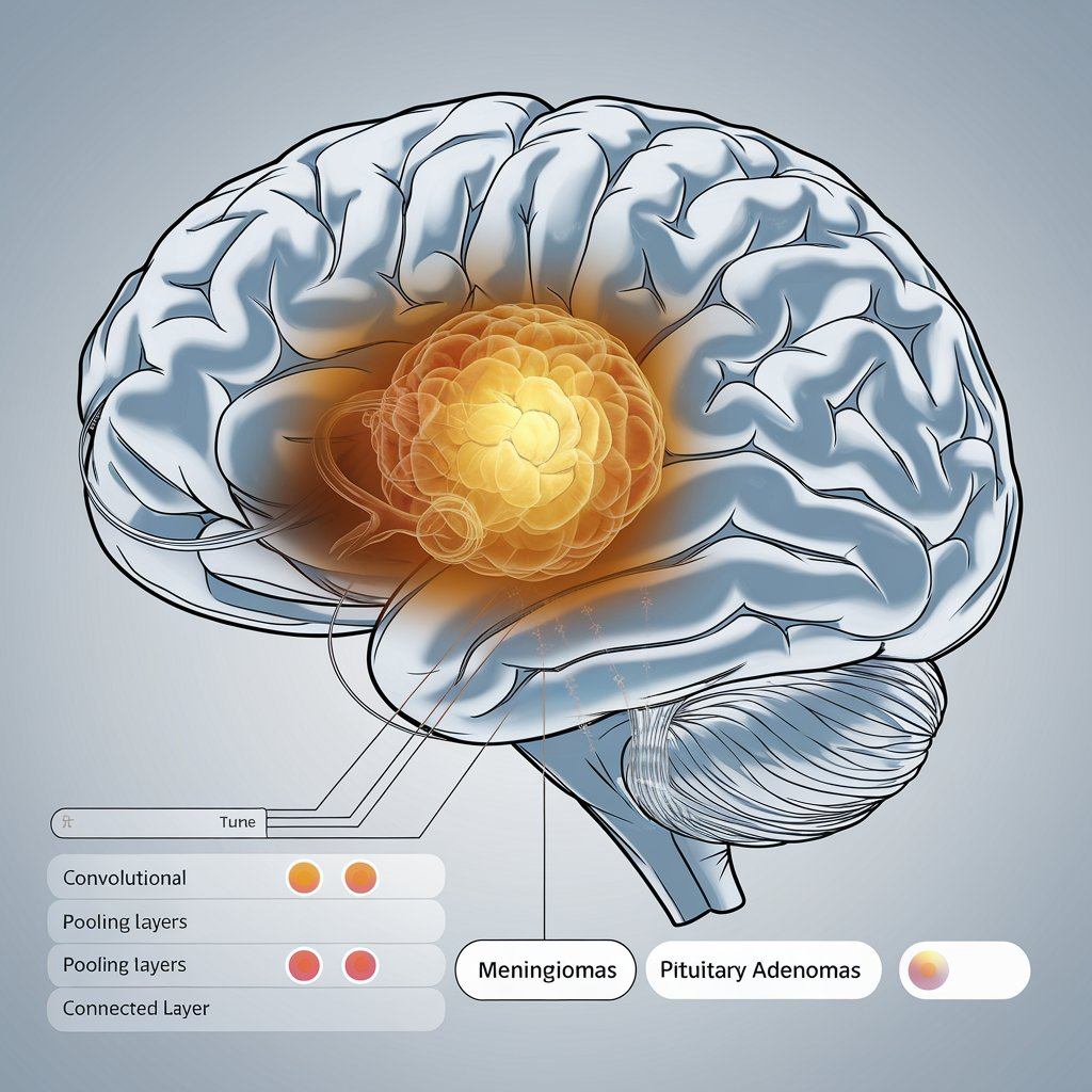
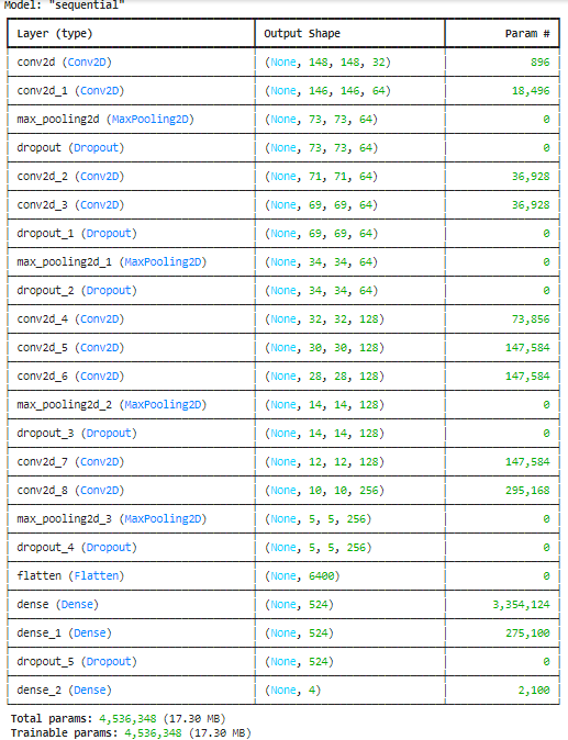
Once the CNN model is trained, it is used to make predictions on unseen MRI images. The model categorizes the brain tumor into one of the four classes: Glioma, Meningioma, Pituitary Tumor, or No Tumor. These predictions are compared to actual labels to evaluate the model's performance.
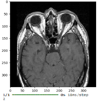
Predicting Image: Shows No tumor
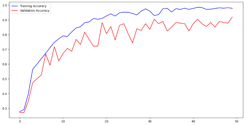
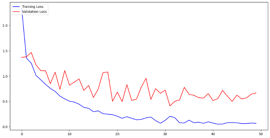
The following graphs show the accuracy and loss over the training and validation datasets during the model's training process:
The accuracy graph illustrates how the model's prediction accuracy improves over time as it learns from the training data.
The loss graph shows the reduction in error (loss) during training and how well the model generalizes to new data.
In medical applications, sensitivity (true positive rate) and specificity (true negative rate) are crucial metrics to ensure that the model can accurately distinguish between tumor types.
AI-based brain tumor classification models are increasingly being integrated into clinical decision support systems (CDSS), assisting radiologists and doctors in diagnosing and planning treatments.
Classified tumor data is being used in research to understand the progression of brain tumors and to develop personalized treatment plans and drugs for patients.
As AI models become more complex, ensuring transparency and interpretability will be key. Explainable AI (XAI) seeks to make AI decisions more understandable for clinicians, enhancing trust and adoption. Future systems will likely integrate various data sources, such as genomic information, clinical data, and imaging scans, to provide a more comprehensive view of brain tumors and improve classification accuracy.

Brain tumor classification using Convolutional Neural Networks (CNNs) offers an effective approach for automating the diagnosis process, improving accuracy, and speeding up detection. By applying advanced AI techniques, radiologists and clinicians can enhance their ability to diagnose and treat brain tumors early, leading to better patient outcomes. Despite challenges such as limited data availability and tumor heterogeneity, ongoing advancements in AI models and data preprocessing techniques promise a brighter future for brain tumor classification. The integration of AI into medical imaging will continue to revolutionize how brain tumors are detected, classified, and treated.
Powered by Froala Editor